Page 14 - Adlershof Special 41
P. 14
Nicht nur die morphologischen Strukturen, auch
die räumliche Verteilung von biologisch wich-
tigen Elementen wie Phosphor oder Schwefel
lassen sich durch deren charakteristische Röntgen-
fluoreszenzstrahlung sichtbar machen. Sogar die
Verteilung chemischer Bindungszustände kann
mittels der hohen Energieauflösung im Röntgen-
mikroskop durch Nahkantenspektroskopie darge-
stellt werden.
Neben den Lebenswissenschaften findet die
Röntgenmikroskopie auch Anwendung in der
Material- und Energieforschung. So untersuch-
ten die Forscher am HZB, in Kooperation mit dem
Institut für Physik der Humboldt-Universität zu
Berlin, Strukturen organischer LEDs. Zurzeit ist
das Röntgenmikroskop aufgrund von Umbau-
arbeiten außer Betrieb. Weitere Kooperationen
mit den Instituten der Humboldt-Universität, wie
dem Integrative Research Institute for the Sciences
(IRIS), sind angedacht. ah Mit Röntgenmikroskopie können z.B. Viren untersucht werden.
X ray microscopes can for one investigate viruses.
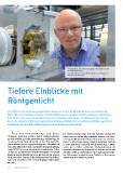
Gerd Schneider, Leiter der Arbeitsgruppe Mikroskopie am HZB
am Röntgenmikroskop.
Gerd Schneider, Head of the Microscopy Research Group at the
HZB at a X ray microscope.
Deeper insights with X rays
Tiefere Einblicke mit Since 2003 the Microscopy Research Group at the Institute of Soft Matter and Functional Mate-
rials of the Helmholtz-Zentrum Berlin (HZB) has been working on high resolution imagery with
Röntgenlicht X rays. Today, one of the world’s most modern X ray microscopes can be found at the electron
storage ring BESSY II in Adlershof. A source of synchrotron radiation, BESSY II generates an intense
bundle of X rays with a physical resolution as low as ten nanometres.
Seit 2003 forscht die Arbeitsgruppe Mikroskopie am Institut für Weiche Materie und funktionale
Materialien des Helmholtz-Zentrums Berlin (HZB) an der hochauflösenden Darstellung mit Röntgen- A lthough electron microscopes can depict even small- Not only the morphological structures, also the spatial dis-
licht. Heute steht eines der weltweit modernsten Röntgenmikroskope am Elektronenspeicherring er structures, X ray microscopes offer many advantages. An tribution of key biological elements like phosphorus or sul-
electron microscope penetrates the sample material only to
phur can be rendered visible in the form of their character-
BESSY II in Adlershof. Mit seiner Synchrotronstrahlung stellt BESSY II eine intensive Röntgenquelle zur a depth less than one micrometre. A typical biological cell, istic X ray fluorescence. The high energy resolution in an X
Verfügung, die räumliche Auflösungen bis zu zehn Nanometern erlaubt. however, is about ten micrometres thick, and the soft X rays ray microscope can even depict the distribution of chemical
generated by BESSY II penetrate whole cells. “Just as com- bonding states thanks to near edge spectroscopy.
puted tomography can depict a head in three dimensions, Not only the life sciences, also materials and energy research
A uch wenn Elektronenmikroskope noch kleinere Im Elektronenmikroskop herrscht Vakuum, biologische we can do the same with the internal structures of a single benefit from X ray microscopy. For instance, researchers at
cell,” explained Gerd Schneider, Head of the Microscopy Re-
Strukturen darstellen können, haben Röntgenmikrosko- Proben müssen aufwendig vorbereitet werden. Zur Unter- search Group at the HZB. the HZB, in cooperation with the Department of Physics at
pe viele Vorteile. Ein Elektronenmikroskop durchdringt das suchung im Röntgenmikroskop wird Gewebe nur schockge- Humboldt-Universität zu Berlin, analysed the structures
zu untersuchende Material nur bis zu eine Tiefe von unter froren. So ist es beispielsweise möglich, bestimmte Proteine An electron microscope contains a vacuum, so biological of organic LEDs. At present, the X ray microscope has been
einem Mikrometer. Eine typische biologische Zelle jedoch ist in einer Zelle durch Farbstoffe im Lichtmikroskop zu lokali- samples must undergo complex preparations. For analyses put out of operation for modifications. Ideas have been put
etwa zehn Mikrometer dick, und weiche Röntgenstrahlung, sieren, um im höher auflösenden Röntgenmikroskop die in an X ray microscope, tissue need only be shock frozen. A forward for further cooperation projects with Humboldt-
so wie BESSY II sie erzeugt, durchleuchtet vollständige Zellen. zellulären Strukturen im Bereich dieses Proteins, an ein und light microscope, for instance, can then locate stains mark- Universität institutes, e.g. the Integrative Research Institute
„So wie ein medizinischer Computertomograph einen Kopf derselben Zelle, zu untersuchen. Diese sogenannte korrela- ing certain proteins in a cell, and a high resolution X ray mi- for the Sciences (IRIS).
dreidimensional abbilden kann, so können wir die inneren tive Mikroskopie ermöglicht neue Einblicke in den inneren croscope can analyse the cellular structure of this protein,
Strukturen einer einzelne Zelle dreidimensional darstellen“, Aufbau und die Funktion von Zellen. both on the one and the same cell. This so called correlative
sagt Gerd Schneider, Leiter der Arbeitsgruppe Mikroskopie microscopy opens up new insights into the internal struc-
am HZB. ture and the function of cells.
14 Adlershof special 41 Adlershof special 41 15
die räumliche Verteilung von biologisch wich-
tigen Elementen wie Phosphor oder Schwefel
lassen sich durch deren charakteristische Röntgen-
fluoreszenzstrahlung sichtbar machen. Sogar die
Verteilung chemischer Bindungszustände kann
mittels der hohen Energieauflösung im Röntgen-
mikroskop durch Nahkantenspektroskopie darge-
stellt werden.
Neben den Lebenswissenschaften findet die
Röntgenmikroskopie auch Anwendung in der
Material- und Energieforschung. So untersuch-
ten die Forscher am HZB, in Kooperation mit dem
Institut für Physik der Humboldt-Universität zu
Berlin, Strukturen organischer LEDs. Zurzeit ist
das Röntgenmikroskop aufgrund von Umbau-
arbeiten außer Betrieb. Weitere Kooperationen
mit den Instituten der Humboldt-Universität, wie
dem Integrative Research Institute for the Sciences
(IRIS), sind angedacht. ah Mit Röntgenmikroskopie können z.B. Viren untersucht werden.
X ray microscopes can for one investigate viruses.
Gerd Schneider, Leiter der Arbeitsgruppe Mikroskopie am HZB
am Röntgenmikroskop.
Gerd Schneider, Head of the Microscopy Research Group at the
HZB at a X ray microscope.
Deeper insights with X rays
Tiefere Einblicke mit Since 2003 the Microscopy Research Group at the Institute of Soft Matter and Functional Mate-
rials of the Helmholtz-Zentrum Berlin (HZB) has been working on high resolution imagery with
Röntgenlicht X rays. Today, one of the world’s most modern X ray microscopes can be found at the electron
storage ring BESSY II in Adlershof. A source of synchrotron radiation, BESSY II generates an intense
bundle of X rays with a physical resolution as low as ten nanometres.
Seit 2003 forscht die Arbeitsgruppe Mikroskopie am Institut für Weiche Materie und funktionale
Materialien des Helmholtz-Zentrums Berlin (HZB) an der hochauflösenden Darstellung mit Röntgen- A lthough electron microscopes can depict even small- Not only the morphological structures, also the spatial dis-
licht. Heute steht eines der weltweit modernsten Röntgenmikroskope am Elektronenspeicherring er structures, X ray microscopes offer many advantages. An tribution of key biological elements like phosphorus or sul-
electron microscope penetrates the sample material only to
phur can be rendered visible in the form of their character-
BESSY II in Adlershof. Mit seiner Synchrotronstrahlung stellt BESSY II eine intensive Röntgenquelle zur a depth less than one micrometre. A typical biological cell, istic X ray fluorescence. The high energy resolution in an X
Verfügung, die räumliche Auflösungen bis zu zehn Nanometern erlaubt. however, is about ten micrometres thick, and the soft X rays ray microscope can even depict the distribution of chemical
generated by BESSY II penetrate whole cells. “Just as com- bonding states thanks to near edge spectroscopy.
puted tomography can depict a head in three dimensions, Not only the life sciences, also materials and energy research
A uch wenn Elektronenmikroskope noch kleinere Im Elektronenmikroskop herrscht Vakuum, biologische we can do the same with the internal structures of a single benefit from X ray microscopy. For instance, researchers at
cell,” explained Gerd Schneider, Head of the Microscopy Re-
Strukturen darstellen können, haben Röntgenmikrosko- Proben müssen aufwendig vorbereitet werden. Zur Unter- search Group at the HZB. the HZB, in cooperation with the Department of Physics at
pe viele Vorteile. Ein Elektronenmikroskop durchdringt das suchung im Röntgenmikroskop wird Gewebe nur schockge- Humboldt-Universität zu Berlin, analysed the structures
zu untersuchende Material nur bis zu eine Tiefe von unter froren. So ist es beispielsweise möglich, bestimmte Proteine An electron microscope contains a vacuum, so biological of organic LEDs. At present, the X ray microscope has been
einem Mikrometer. Eine typische biologische Zelle jedoch ist in einer Zelle durch Farbstoffe im Lichtmikroskop zu lokali- samples must undergo complex preparations. For analyses put out of operation for modifications. Ideas have been put
etwa zehn Mikrometer dick, und weiche Röntgenstrahlung, sieren, um im höher auflösenden Röntgenmikroskop die in an X ray microscope, tissue need only be shock frozen. A forward for further cooperation projects with Humboldt-
so wie BESSY II sie erzeugt, durchleuchtet vollständige Zellen. zellulären Strukturen im Bereich dieses Proteins, an ein und light microscope, for instance, can then locate stains mark- Universität institutes, e.g. the Integrative Research Institute
„So wie ein medizinischer Computertomograph einen Kopf derselben Zelle, zu untersuchen. Diese sogenannte korrela- ing certain proteins in a cell, and a high resolution X ray mi- for the Sciences (IRIS).
dreidimensional abbilden kann, so können wir die inneren tive Mikroskopie ermöglicht neue Einblicke in den inneren croscope can analyse the cellular structure of this protein,
Strukturen einer einzelne Zelle dreidimensional darstellen“, Aufbau und die Funktion von Zellen. both on the one and the same cell. This so called correlative
sagt Gerd Schneider, Leiter der Arbeitsgruppe Mikroskopie microscopy opens up new insights into the internal struc-
am HZB. ture and the function of cells.
14 Adlershof special 41 Adlershof special 41 15